Many small structures within the body are difficult or impossible to visualize using external imaging methods. Endoscopy has long been the preferred technique, when feasible, for observing such structures. Thinner endoscopes can access more regions with minimal disruption to surrounding tissue. However, the benefits of thinner endoscopes are often counterbalanced by imaging challenges. Optical fields relayed through each fiber core can be significantly distorted due to phase scrambling caused by surface irregularities at the fiber ends or the presence of multi-mode cores. Additionally, achieving high-quality endoscopic images typically requires the fiber tip to be positioned close to the target or necessitates the integration of a lens. Imaging structured in 3D or through scattering layers beyond the fiber tip is often not feasible with conventional methods.
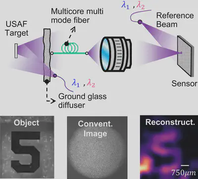
In this project, we address these limitations by integrating Synthetic Wavelength Imaging with fiber endoscopy. The resulting system eliminates the need for lenses and is inherently resilient to phase scrambling introduced by scattering and fiber bending. Using this approach, we have successfully demonstrated the endoscopic imaging of features approximately 750 micrometers in size on objects located behind a scattering layer. This advancement opens new possibilities for achieving spatially resolved three-dimensional imaging of structures concealed beneath tissue, significantly expanding the potential of fiber endoscopes for biomedical applications.