Medical Applications of 3D Surface Measurement
Accurate, robust, and fast 3D surface measurements are crucial in modern medical imaging and can support a wide range of clinical applications from surgical guidance to telemedicine. For instance, providing surgeons with an accurate, real-time 3D view of the surgical field can be essential for complex procedures, e.g., in robotic-assisted or AR/VR-assisted surgeries. However, established techniques like structured light triangulation can face significant difficulties in surgical environments due to partially specular reflections from wet, bloody surfaces. Several of our developments aim to actively address these key challenges that have historically limited the use of advanced 3D surface measurement techniques in medical settings. Moreover, we are working on novel methods to enable high-quality 3D imaging using commodity devices like tablets and smartphones with the goal to make basic diagnostic tools, e.g., for the 3D analysis of skin lesions, more accessible. Additionally, our previous projects have focused on seamless co-registration of multimodal imaging data (MRI, EEG, MEG) and motion compensation during procedures like radiation therapy or CT imaging, leveraging real-time 3D sensing for improved results.
Besides imaging the surface of the human body, a significant part of our research portfolio is devoted to imaging through scattering media, which has potential applications in medical imaging to image deep inside the human tissue (see our Synthetic Wavelength Imaging research track for more info).
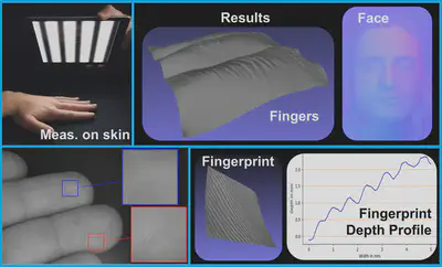
SkinScan
Selected Publications
Skinscan: Low-cost 3d-scanning for dermatologic diagnosis and documentation.
2021 IEEE International Conference on Image Processing (ICIP), 2021.
Skinscan: 3D Dermatologic Diagnosis and Documentation with Commodity Devices.
2021 55th Asilomar Conference on Signals, Systems, and Computers, 2022.
Single-Shot Fringe Projection Triangulation on Wet and Shiny tissue

Selected Publications
Radiometric calibration of active 3D imaging setups using superquadric fitting.
Interferometry and Structured Light 2024, 2024.
“On-the-Fly” Radiometric Calibration of Active 3D Imaging Setups using Superellipse Fitting.
Frontiers in Optics, 2023.
Hand-Guided Single-Shot Multi-Line Triangulation
Visit the Osmin3D YouTube channel for more Videos.
Color 3D Movie of a talking face - RAW data (no post processing)
Color 3D Movie of another talking face - RAW data (no post processing)
Real-time 3D movie of a boncing ping-pong ball - RAW data (no post processing)
Real-time 3D movie of a folded paper - RAW data (no post processing). High object frequencies are preserved
How to watch a ‘3D movie’
3D movie of a talking face with unidirectional lines plus closeup - RAW data (no post processing).
3D movie of a talking face with unidirectional lines - RAW data (no post processing).
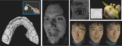
Flying Triangulation Dental Scanner
Flying Triangulation Face Scanner
3D models measured with Flying Triangulation (no post processing).
360° scan of a plaster bust.
Selected Publications
Frontiers in neuroscience 8, 42, 2014.
Management of head motion during MEG recordings with Flying Triangulation.
Proceedings of the DGaO Conference, 2013.
Flying Triangulation: a tool for automated medical diagnosis.
Proceedings of the DGaO Conference, 2014.
Improved EEG source localization employing 3D sensing by\" Flying Triangulation\".
Videometrics, Range Imaging, and Applications XII; and Automated Visual Inspection, 2013.
Sparse Active Triangulation Grids for Respiratory Motion Management.
Deutsche Gesellschaft für angewandte Optik (DGaO), 2012.
Single-Shot 3D Sensing Close to Physical Limits and Information Limits.
Book, Springer Theses, 2019, Dissertation, University Erlangen-Nuremberg, 2017.